Home » Without Label » Diagram Of The Muscles In The Forearm / Solved Label Each Of The Indicated Muscles That Move The Forearm Chegg Com - These do not include the hip, neck and forearm muscles, which are rarely trained in isolation.
Diagram Of The Muscles In The Forearm / Solved Label Each Of The Indicated Muscles That Move The Forearm Chegg Com - These do not include the hip, neck and forearm muscles, which are rarely trained in isolation.
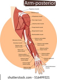
Diagram Of The Muscles In The Forearm / Solved Label Each Of The Indicated Muscles That Move The Forearm Chegg Com - These do not include the hip, neck and forearm muscles, which are rarely trained in isolation.. The pronator teres is a muscle (located mainly in the forearm) that, along with the pronator quadratus, serves to pronate the forearm (turning it so that the palm faces posteriorly when from the anatomical position). For example, contraction of the biceps muscle, attached to the scapula and radius, will raise the forearm. Jun 18, 2018 · the muscles in this area are mostly involved with flexion of your wrist and fingers as well as rotation of your forearm. The sequence shown above is one possible way to order the exercises. These muscles are grouped into the muscles of the thoracic cage and the muscles of the abdominal wall.
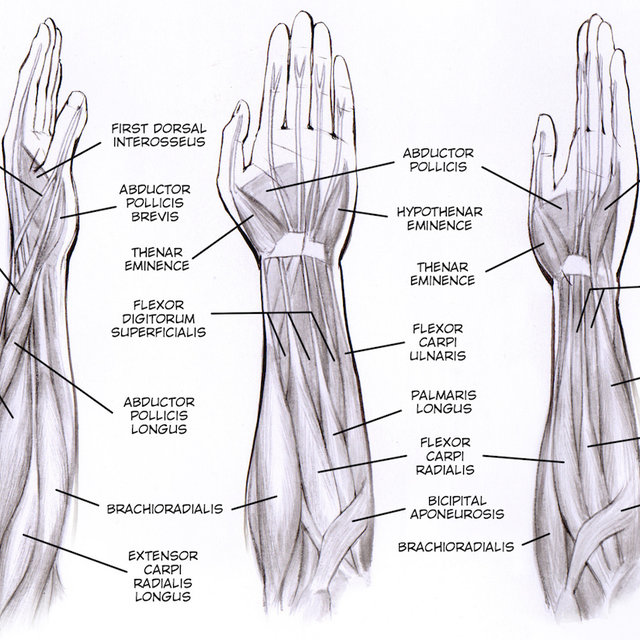
The sequence shown above is one possible way to order the exercises. Bodybuilders commonly divide the body's individual muscles into ten major muscle groups. As seen in this forearm muscles diagram, the flexor muscles reside in the anterior compartment of the forearm, and are separated into the three following layers: These do not include the hip, neck and forearm muscles, which are rarely trained in isolation. Flexor carpi ulnaris, palmaris longus, flexor carpi radialis, and pronator teres.
Forearm Muscles Bones And Anatomy Guide from criticalbody.com Are you feeling overwhelmed by all of the information in this article? There are three planes commonly used; As seen in this forearm muscles diagram, the flexor muscles reside in the anterior compartment of the forearm, and are separated into the three following layers: The most common exercises for these muscle groups are listed above. Jun 18, 2018 · the muscles in this area are mostly involved with flexion of your wrist and fingers as well as rotation of your forearm. May 31, 2021 · diagram of the forearm flexors. The sequence shown above is one possible way to order the exercises. Aug 27, 2018 · anatomy and function of forearm muscles the forearm contains more muscles than the upper arm does.
As seen in this forearm muscles diagram, the flexor muscles reside in the anterior compartment of the forearm, and are separated into the three following layers:
It contains both an anterior and posterior compartment, and each is further divided into layers. For example, contraction of the biceps muscle, attached to the scapula and radius, will raise the forearm. The ulna is the median bone in the forearm that runs parallel to the radius. Jun 18, 2018 · the muscles in this area are mostly involved with flexion of your wrist and fingers as well as rotation of your forearm. It is one of the five main nerves originating from the brachial plexus. The sequence shown above is one possible way to order the exercises. May 31, 2021 · diagram of the forearm flexors. Are you feeling overwhelmed by all of the information in this article? Superficial layer flexor carpi ulnaris. As seen in this forearm muscles diagram, the flexor muscles reside in the anterior compartment of the forearm, and are separated into the three following layers: The most common exercises for these muscle groups are listed above. There are three planes commonly used; These muscles are grouped into the muscles of the thoracic cage and the muscles of the abdominal wall.
It is one of the five main nerves originating from the brachial plexus. Jun 18, 2018 · the muscles in this area are mostly involved with flexion of your wrist and fingers as well as rotation of your forearm. The most common exercises for these muscle groups are listed above. There are three planes commonly used; Jul 27, 2021 · anterolateral trunk muscles diagram the anterior trunk muscles cover the anterolateral part of the trunk by attaching to the bony framework of the thoracic cage and pelvis.
Arm Muscle Anatomy High Res Stock Images Shutterstock from image.shutterstock.com Are you feeling overwhelmed by all of the information in this article? The most common exercises for these muscle groups are listed above. These muscles are grouped into the muscles of the thoracic cage and the muscles of the abdominal wall. Jul 27, 2021 · anterolateral trunk muscles diagram the anterior trunk muscles cover the anterolateral part of the trunk by attaching to the bony framework of the thoracic cage and pelvis. These do not include the hip, neck and forearm muscles, which are rarely trained in isolation. The sequence shown above is one possible way to order the exercises. The pronator teres is a muscle (located mainly in the forearm) that, along with the pronator quadratus, serves to pronate the forearm (turning it so that the palm faces posteriorly when from the anatomical position). As seen in this forearm muscles diagram, the flexor muscles reside in the anterior compartment of the forearm, and are separated into the three following layers:
Bodybuilders commonly divide the body's individual muscles into ten major muscle groups.
The ulna is the median bone in the forearm that runs parallel to the radius. Are you feeling overwhelmed by all of the information in this article? The sequence shown above is one possible way to order the exercises. Flexor carpi ulnaris, palmaris longus, flexor carpi radialis, and pronator teres. As seen in this forearm muscles diagram, the flexor muscles reside in the anterior compartment of the forearm, and are separated into the three following layers: It contains both an anterior and posterior compartment, and each is further divided into layers. The median nerve is a nerve in humans and other animals in the upper limb. Aug 27, 2018 · anatomy and function of forearm muscles the forearm contains more muscles than the upper arm does. It is one of the five main nerves originating from the brachial plexus. The pronator teres is a muscle (located mainly in the forearm) that, along with the pronator quadratus, serves to pronate the forearm (turning it so that the palm faces posteriorly when from the anatomical position). Superficial layer flexor carpi ulnaris. The most common exercises for these muscle groups are listed above. These do not include the hip, neck and forearm muscles, which are rarely trained in isolation.
Flexor carpi ulnaris, palmaris longus, flexor carpi radialis, and pronator teres. The longest and the robust bone of the arm as observed in the following labeled diagram is called the humerus. The median nerve is a nerve in humans and other animals in the upper limb. These do not include the hip, neck and forearm muscles, which are rarely trained in isolation. These muscles are grouped into the muscles of the thoracic cage and the muscles of the abdominal wall.
Forearm Muscle Diagram A Contrast Sketch Of Forearm Muscles With The Download Scientific Diagram from www.researchgate.net Bodybuilders commonly divide the body's individual muscles into ten major muscle groups. The ulna is the median bone in the forearm that runs parallel to the radius. Are you feeling overwhelmed by all of the information in this article? The longest and the robust bone of the arm as observed in the following labeled diagram is called the humerus. These do not include the hip, neck and forearm muscles, which are rarely trained in isolation. Superficial layer flexor carpi ulnaris. It contains both an anterior and posterior compartment, and each is further divided into layers. There are three planes commonly used;
Jan 06, 2018 · using anatomical planes allows for accurate description of a location, and also allows the reader to understand what a diagram or picture is trying to show.
Jul 27, 2021 · anterolateral trunk muscles diagram the anterior trunk muscles cover the anterolateral part of the trunk by attaching to the bony framework of the thoracic cage and pelvis. These do not include the hip, neck and forearm muscles, which are rarely trained in isolation. May 31, 2021 · diagram of the forearm flexors. Flexor carpi ulnaris, palmaris longus, flexor carpi radialis, and pronator teres. It is one of the five main nerves originating from the brachial plexus. The most common exercises for these muscle groups are listed above. The pronator teres is a muscle (located mainly in the forearm) that, along with the pronator quadratus, serves to pronate the forearm (turning it so that the palm faces posteriorly when from the anatomical position). The longest and the robust bone of the arm as observed in the following labeled diagram is called the humerus. The median nerve is a nerve in humans and other animals in the upper limb. Jan 06, 2018 · using anatomical planes allows for accurate description of a location, and also allows the reader to understand what a diagram or picture is trying to show. The sequence shown above is one possible way to order the exercises. As seen in this forearm muscles diagram, the flexor muscles reside in the anterior compartment of the forearm, and are separated into the three following layers: Superficial layer flexor carpi ulnaris.